SmartIR reduces waste, increases yields, and improves output quality, especially as organizations migrate towards more autonomous operations in manufacturing. A valuable quality assurance tool, SmartIR is able to distinguish minute variations in manufacturing outputs to ensure consistent production.

 Multifaceted Analytics & Customizable Dashboards
SmartIR provides deep analytics and easy-to-digest dashboards to inform your predictive maintenance and facility safety processes
 Real-time, Automated Notifications
Instant alters communicate imminent maintenance needs or safety concerns; tiered communication options ensure the right team members are immediately notified.
 Seamless Integration with VMS and DCS Systems
Seamlessly integrate into organizational systems and processes, including VMS and DCS platforms.
 On-Site or Remote Video Monitoring
Access real-time or archived video feeds from the thermal cameras, anywhere with an internet connection.


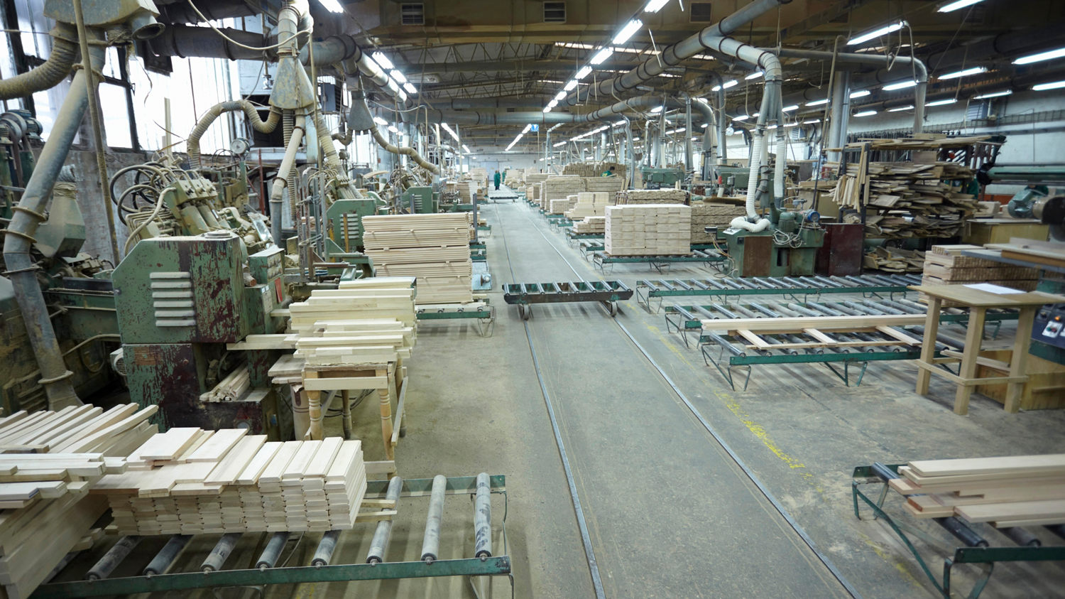
Case Study: Early Fire Prevention in Saw Mill
After a fire caused a sawmill shutdown, a large pulp and paper...
Read more
#34: AWS Innovation with ICI
Listen on your preferred podcast platform: Spotify, Apple Podcasts, Google...
Read more
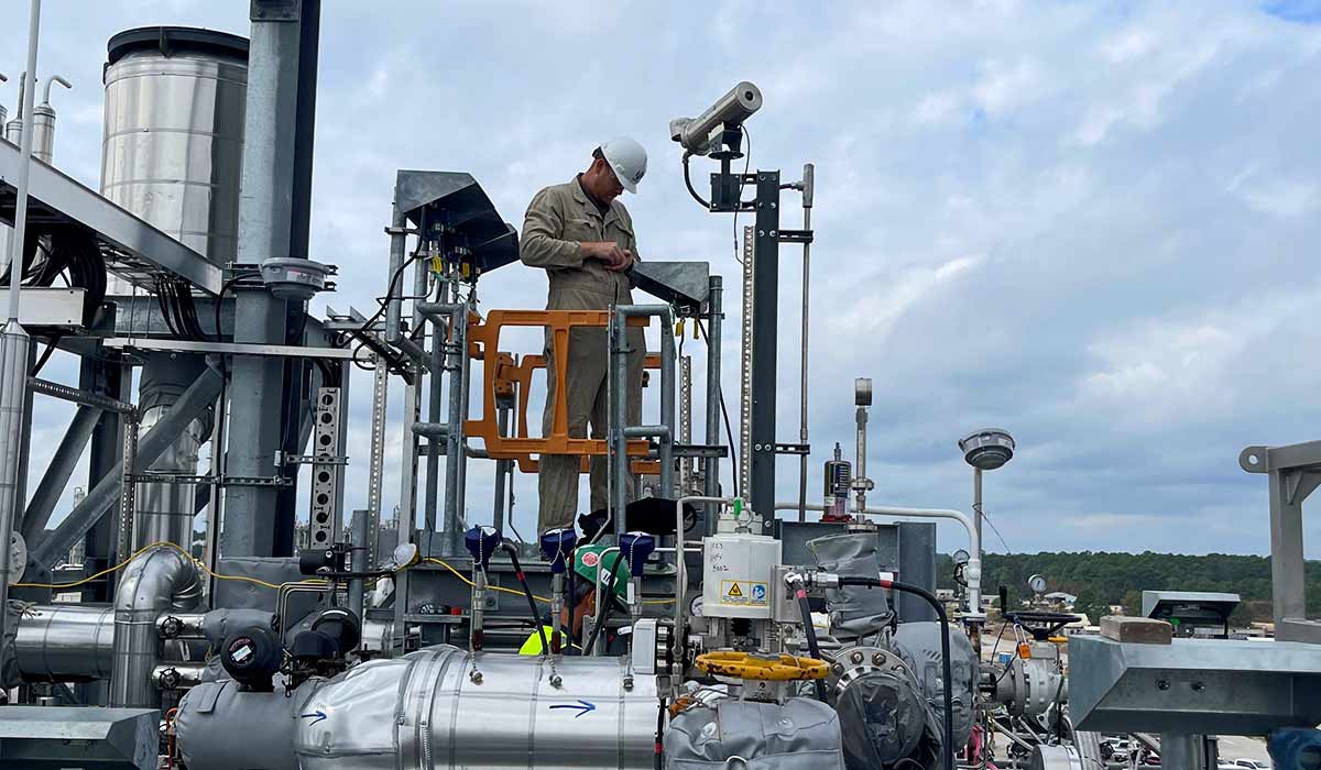
How Optical Gas Imaging Can Improve Plant Safety
If you’re a plant safety manager, you need accurate and actionable...
Read more
